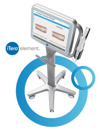
iTero Element Scanner
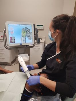
During the impression process, you can breathe or swallow as you normally would. You can even pause during the process if you need to sneeze or just want to ask a question. The scanner gives us a 3D model of your mouth that can be used for your dental services including the Invislaign Outcome Simulator to show you what your new smile might look like. 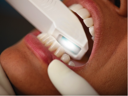
|
We Are a Digital Impression Practice. Proud Owners of an iTero Element Intraoral Scanner
We'll Make a Great First Impression The iTero Element Scanner is a state-of-the-art digital impression system that eliminates the need for messy putty in your mouth. With our iTero Element Scanner, we can digitally capture a detailed 3D model of your teeth and gums. Not only is this process far more comfortable than the old putty based impressions, but it's faster and can offer a superior clinical endpoint.
|
2D & 3D Cone Beam Digital Panoramic Machine
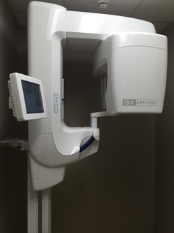
The GXDP-700 Series allows different types of imaging for a wide range of clinical situations, giving us the flexibility to capture the images necessary for the procedures we perform. From general preventative care to implants, T.M.J (tempromandibular joint) and extractions.
The Safety of Dental X-Rays
Both dental professionals and dental patients have probably wondered about the safety of dental x-rays at least once. Many studies have been done on the effects of both medical and dental x-rays on patient health.
What the Research Says
According to the American College of Radiology, four bite wing x-rays expose a patient to approximately the same amount of radiation that the patient receives from the sun and other sources each day.
Digital x-ray machines generally produce half the amount of radiation that film based x-rays do
When performed correctly and with proper safety procedures, dental x-rays pose a very small risk to patients.
When is it Safe to be Exposed to X-Rays?
The ionizing radiation that you receive from one dental x-ray is substantially less than the radiation you receive every day from the sun and stars. Advances in technology have made dental x-rays much safer. Doses of radiation are kept at the lowest practical value to minimize patient exposure. This is done with the use of a long cone position-indicating device, appropriate setting on the machine.
Digital Radiography
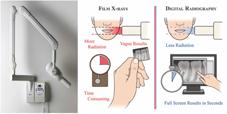
Digital radiography is a form of x-ray imaging, where digital X-ray sensors are used instead of traditional photographic film. Advantages include time efficiency through bypassing chemical processing and the ability to digitally transfer and enhance images. Also less radiation can be used to produce an image of similar contrast to conventional radiography.
Instead of X-ray film, digital radiography uses a digital image capture device. This gives advantages of immediate image preview and availability; elimination of costly film processing steps; a wider dynamic range, which makes it more forgiving for over- and under-exposure; as well as the ability to apply special image processing techniques that enhance overall display of the image.
Instead of X-ray film, digital radiography uses a digital image capture device. This gives advantages of immediate image preview and availability; elimination of costly film processing steps; a wider dynamic range, which makes it more forgiving for over- and under-exposure; as well as the ability to apply special image processing techniques that enhance overall display of the image.
Sterilization
We use extensive sterilization procedures, going well beyond the guidelines recommended by the ADA and CDC. We use barrier techniques such as disposable masks and gloves and replace them with every new patient. Each piece of fixed operatory equipment is wiped-down with antibacterial/antiviral solution prior to every clinical procedure and plastic barriers placed between patients. We use autoclave sterilization for all instruments prior to dental examination and treatment, in addition, we use a biological monitoring service daily to ensure constant sterilization.
You can be assured that our staff have been specifically trained on sterilization procedures and it’s importance.
You can be assured that our staff have been specifically trained on sterilization procedures and it’s importance.